Laboratory for Evolutionary Cell Biology of the Skin
School of Bioscience and Biotechnology
Tokyo University of Technology
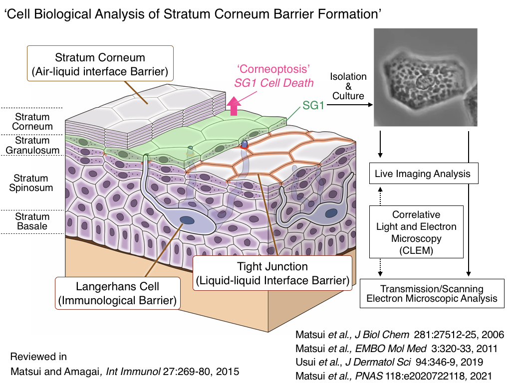
Skin epidermis is the outer most barrier of our body. The skin surface is composed of dead stratum corneum which serves as air-liquid interface barrier. To understand the formation of this functional barrier, we focus to the final living layer, stratum granulosum cells (SG1). In combination with light and electron microscopic techniques, we are dissecting how the SG1 cells die termed ‘corneoptosis’ and form SC during cornification process. Particularly, we will study comparative evolutionary cell biology of SG1 cells in various terrestrial vertebrates (amphibians, reptiles, aves and mammals) to understand the adaptive evolution of epithelial cells.
News and Topics
- 2025/4/30 We started Instagram.

- On May 15, 2024, a research study conducted by an international collaborative team, including Professor Takeshi Matsui (Tokyo University of Technology, formerly at RIKEN), the Department of Dermatology at Keio University School of Medicine, and the Laboratory for Skin Homeostasis at RIKEN Center for Integrative Medical Sciences (both are led by Professor Masayuki Amagai), was published online in Nature Communications.
The study reveals that the outermost layer of the skin, the stratum corneum, which plays a critical role in the skin barrier function, forms a three-layered pH (hydrogen ion concentration) structure. This previously unrecognized pH stratification is essential for maintaining the homeostasis of the stratum corneum. This work was initiated by Yuki Furuichi, who was a graduate student at the time.
Fukuda K, Ito Y, Furuichi Y, Matsui T, Horikawa H, Miyano T, Okada T, Logtestijn V. M, Tanaka J. R, Miyawaki A, Amagai M:Three stepwise pH progressions in stratum corneum for homeostatic maintenance of the skin. Nature Communications 15:4062, 2024.
- 2022/5/17 We reported a new staining method for electron microscope specimens. The method is based on double staining with hematoxylin, which is widely used as a staining agent for light microscopy, and lead solution. It is expected to replace the conventional method of double staining with uranium acetate and lead solution and is a better choice in terms of safety, cost, and ease of handling. The research results were published in the online edition of Scientific Reports.
Sasaki H, Arai H, Kikuchi E, Saito H, Seki K, Matsui T: Novel electron microscopic staining method using traditional dye, hematoxylin. Sci Rep. 12:7756, 2022.
Press Release: Development of a new staining method for electron microscope specimens –Expected to be a safe, cost-effective, and easy-to-use method–